
Steatocystoma Multiplex: What It Is, Symptoms, Diagnosis and All Treatment Options
Steatocystoma multiplex is a benign cyst under the skin that arises from the duct of the oil gland (sebaceous duct) and contains clear, odorless oil. It usually appears as many bumps at once on the chest, arms, armpits and neck. It does not go away on its own, does not turn into cancer, and can be treated by removing the cyst wall.
How Steatocystoma Multiplex Differs from Epidermoid Cyst and Pilar Cyst
These three kinds of lumps under the skin look very similar, but their origin and contents are clearly different, which determines the treatment and the chance of recurrence.
Item | Steatocystoma Multiplex | Epidermoid cyst | Pilar cyst (Trichilemmal cyst) |
|---|---|---|---|
Origin | Duct of the oil gland (sebaceous duct) | Epidermal skin cells that have become embedded in the dermis | Outer root sheath of the hair follicle |
Contents | Clear, odorless oil | Thick, white, cream-like keratin with a characteristic smell | Firm, dense keratin, usually odorless |
Common sites | Chest, arms, armpits, neck | Face, neck, trunk, back | Mostly the scalp |
Number of lumps | Often many bumps at once | Often a single bump or only a few | Single or multiple; can run in families |
Central opening (punctum) | Usually absent | A dark central opening is often visible | Usually absent |
Recurrence after the wall is fully removed | The same lump usually does not return, but new bumps may appear elsewhere because of a genetic tendency | Low, if the cyst wall is removed completely | Low; usually comes out as a single piece |
Main treatment | CO2 laser, RF microneedle, minor surgery | Surgical removal of the entire cyst sac | Surgical removal of the entire cyst sac |
The quickest way to tell them apart in the clinic is what is inside the lump — if puncturing it gives clear, odorless oil, it is usually steatocystoma; if it gives thick, white, cream-like material with a smell, it is usually an epidermoid cyst; and if it is a lump on the scalp that comes out as a whole sac, it is usually a pilar cyst.
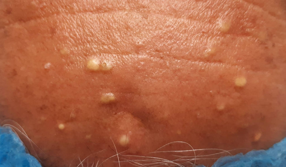
Steatocystoma multiplex cysts are a type of skin cyst that often worries patients because many bumps appear at once, spread across the trunk, arms, legs or the middle of the chest. Although it is a harmless growth and almost never becomes cancerous, it can affect confidence and may become inflamed and pus-filled. In this article, the dermatology specialist team at Siam Dermatology (a specialist skin clinic) covers it in full, from how it forms and how to tell it apart from similar-looking cysts, to every treatment with the advantages and limitations of each.
What Is Steatocystoma Multiplex?
Steatocystoma multiplex is a cyst that forms in the duct of the oil gland (sebaceous duct). The cyst wall looks similar to that of an epidermal cyst, but the contents differ: in steatocystoma they are oil or fat produced by the oil glands, not keratin. When punctured, a yellow, butter-like oil flows out, a characteristic feature that helps with diagnosis.
The word "multiplex" means that it usually appears as many bumps at once, from a few to dozens, unlike steatocystoma simplex, which is a single bump.
Causes and Genetic Inheritance
Steatocystoma multiplex is in part inherited in an autosomal dominant pattern, linked to mutations in the keratin 17 (KRT17) gene, the same gene found in pachyonychia congenita syndrome, so some families have several affected members. However, a number of patients develop it on their own with no family history (sporadic). It usually begins between the teenage years and early adulthood, when the oil glands become more active because of hormones.
Symptoms and Signs to Look For
Bumps or lumps under the skin, from 2–3 mm up to about 5 cm (most are no larger than 5 cm)
Many bumps, appearing in clusters
Common sites: the middle of the chest, upper arms, thighs, armpits, neck and trunk
Shallow cysts look like yellow bumps; deeper ones are the same color as the skin
Soft to fairly firm to the touch, slightly movable, and mostly painless
When punctured, yellow oil flows out (a characteristic feature)
Some bumps may become inflamed, red, painful and pus-filled, especially in areas with frequent friction. This is called steatocystoma multiplex suppurativa, and it may leave scars after it heals
Differential Diagnosis: How Steatocystoma Differs from Other Lumps
Many people mistake it for acne or an ordinary fatty lump. A dermatology specialist tells it apart from similar-looking conditions as follows.
Steatocystoma multiplex: the contents are liquid oil or fat; usually many bumps; puncturing gives yellow oil; thin wall
Epidermoid cyst: the contents are white, flaky keratin with a smell; usually a single bump with a central opening (punctum)
Milia: keratin; very small, shallow white bumps, usually on the face
Lipoma: true fatty tissue; soft, large, deep lump that rolls under the fingers
Clogged pores / inflamed acne: related to the hair follicle; has a head; changes quickly
Diagnosis relies mainly on the clinical appearance. When it is unclear, the doctor may use skin ultrasound or take a tissue sample (biopsy) for pathology to confirm.
Does It Need Treatment?
Because steatocystoma is a growth with a very low chance of becoming cancerous, not every lump needs to be removed. Treatment is considered when:
You are concerned about appearance
Lumps are often inflamed or infected, or repeatedly form pus
They are in an area with friction and cause pain
A lump grows abnormally or another condition is suspected
Important to understand before treatment: every method carries a chance of scarring, and treatment does not prevent new lumps from forming, because the condition has a genetic tendency. Planning together with a dermatology specialist is therefore important.
Treatment Options for Steatocystoma Multiplex (All Methods)
1. Carbon Dioxide Laser (CO2 Laser)
Suitable for small to medium cysts. The doctor uses the laser to make a small opening in the skin, then drains the contents and removes the cyst wall. The advantages are a small wound and quick recovery, which suits cases with many bumps. The point to watch is technique: the cyst wall must be removed completely to reduce recurrence.
2. RF Microneedle (Insulated Radiofrequency)
Radiofrequency energy is delivered through needles to destroy the cyst wall from the inside. Suitable for small bumps, with a very small wound, and suitable for people who are concerned about scarring.
3. Minor Surgery (Surgical Excision)
This gives the most permanent result for the lump treated, because both the cyst and its wall are removed. Suitable for lumps that are often inflamed or infected, large lumps, or lumps in areas with friction. The limitation is stitches and more scarring than other methods, so it is usually done only for problem lumps.
4. Needle Aspiration and Wall Removal (Needle Aspiration / Incision & Drainage)
Because the contents are oil, a needle may be used to draw the oil out, followed by removal of the cyst wall. This leaves a small wound, but if the wall is not removed completely, the cyst may come back.
5. Medication (for Inflamed Cysts)
Oral isotretinoin is used for severely inflamed lumps and temporarily reduces inflammation, but the effect often does not last and the lumps return after the medication is stopped.
Antibiotics such as oral tetracycline, topical clindamycin or benzoyl peroxide wash help control inflammation and infection.
If there is pus, the lump must be cut open to drain it, together with antibiotics.
Note: medication controls inflammation but does not remove the cyst. Permanent removal requires a procedure that takes out the cyst wall.
Aftercare and Preventing Scars
Keep the wound clean and dry as your doctor advises
Avoid strong sunlight on the wound area to reduce dark marks, and apply sunscreen once the wound has closed
Do not pick at or squeeze the lumps yourself, as this risks inflammation and scarring
Follow up with your doctor and plan how to manage new lumps that may appear in future
Chance of Recurrence
Because steatocystoma multiplex has a genetic tendency, treatment removes only the lumps that were treated, but new lumps may appear in other places. A good approach is long-term, continuing care with a specialist clinic, so that new lumps are managed early and inflammation is reduced.
When to See a Dermatology Specialist
You should consult a doctor if: a lump is inflamed, red, painful or pus-filled / a lump grows unusually fast / you have many bumps and they affect your confidence / you are not sure which type of cyst it is. A correct diagnosis from the start helps choose the right treatment and reduces the chance of scarring.
Treating Steatocystoma Multiplex at Siam Dermatology
Siam Dermatology (a specialist skin clinic) is run by a team of dermatology specialists experienced with cysts and complex skin lumps. We choose the treatment to suit the size, location and number of lumps for each person, for good results and the least possible scarring.
Consultation / appointments: call +66 61 448 7000 | LINE: @siamderm
Note: results may vary from person to person. This information is for education and is not a substitute for a diagnosis by a doctor.
Frequently Asked Questions (FAQ)
Is steatocystoma multiplex dangerous?
No. It is a benign growth and almost never becomes cancerous, but some bumps may become inflamed and pus-filled, so it should be treated when symptoms appear.
Does this cyst go away on its own?
Generally no, and it usually stays until it is treated. Treatment removes the lumps that were treated but does not prevent new ones.
Can I squeeze it myself?
No, because it risks infection and scarring, and the cyst wall is usually not removed completely, so it comes back.
Does it come back after treatment?
A lump whose wall is completely removed usually does not return in the same place, but because the condition is genetic, new lumps may appear in other places.
Which method leaves the least scarring?
For small bumps, CO2 laser and RF microneedle usually leave smaller wounds than surgery, but the choice depends on size, location and number of lumps, so it should be assessed by a doctor.
How does steatocystoma differ from acne or a fatty lump?
The difference is the contents. Steatocystoma contains oil, while acne and epidermoid cysts contain keratin, and a fatty lump (lipoma) is fatty tissue in a deeper layer. A dermatology specialist can tell them apart from the clinical appearance.
References
DermNet NZ. Steatocystoma multiplex. https://dermnetnz.org/topics/steatocystoma-multiplex
DermNet NZ. Epidermoid cyst. https://dermnetnz.org/topics/epidermoid-cyst
DermNet NZ. Trichilemmal cyst (pilar cyst). https://dermnetnz.org/topics/trichilemmal-cyst
Bolognia JL, Schaffer JV, Cerroni L, eds. Dermatology. 4th ed. Elsevier; 2018 (Cysts of the skin).
UpToDate. Overview of benign lesions of the skin. https://www.uptodate.com
Written by Dr. Patara Voravutinon, Dermatologist · Last reviewed July 30, 2026

Comments